Molecular Visualization: Mass Spec Imaging Delivers Insights for Cancer Research
How does mass spectrometry (MS) imaging provide a molecular image that can be used for the biochemical characterization of histological images?
A rapidly developing technique for clinical research, MS imaging enables investigation of the spatial distribution of molecules at complex surfaces. It’s a direct-from-sample analytical technique that provides researchers with the information needed to quickly and objectively interpret molecular profiles and make confident decisions based on definitive data.
Modern technology for advanced MS imaging includes Matrix Assisted Laser Desorption Ionization (MALDI) and Desorption Electrospray Ionization (DESI), and ion mobility. They provide comprehensive molecular distribution information in applications such as proteomics, metabolomics, cell and tissue biology, research pathology and histology.
This year in our Meet the Expert series at AACR 2016, April 17-20 in New Orleans, we are delighted to have Dr. Arash Zarrine Afsar join us and present the exciting research he is conducting with his team at University Health Network (UHN), Toronto. Leveraging the power of MS imaging, his team is examining biological tissues to both differentiate tumor tissue from healthy tissue, as well as to study tumor heterogeneity.
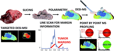
In a recent Chemical Science Journal article, Dr. Afsar’s team demonstrated the use of polarimetry coupled with molecular imaging to accelerate pathological assessment of cancer tissue. They showed that the combination of polarimetry and DESI MS imaging provided fast and accurate pathology assessment for cancer typification in less than 2 minutes – compared to 30 minutes for histopathology of ex vivo tissue slices – without any additional sample preparation.
In addition to his work with polarimetry, Dr. Afsar has also studied the utility of a mapping contrast agent used in magnetic resonance imaging (MRI) agents to target diseased sites—which would reveal tumor heterogeneity in the absence of known mass spectrometry profiles. Through this technique, tumor margins of human breast cancer tumors grown in mice were identified by DESI-MS in corroboration with in vivo MRI results.
If you are attending AACR in New Orleans this year, be sure to stop by the Waters booth during our Meet the Expert series. There, you’ll be able to personally speak with Dr. Arash Zarrine Afsar and our other experts about the analytical techniques used in cancer research.
Learn more:
- Waters at AACR 2016
- Details about our AACR Meet the Expert series in SelectScience
- Full Spectrum Molecular Imaging (DESI and MALDI)
- MS imaging in the health sciences, including cancer research
Related posts

How Improved Selectivity in Immunosuppressant Analysis Can Help Organ Transplant Patients
The Waters MassTrak Immunosuppressant Calibrator, Control, and Internal Standard Sets, soon available, are developed to help laboratories utilize LC-MS/MS with confidence in their assay results.

The Future Generation of LC-MS/MS IVD Solutions
Our MassTrak IVD solutions provide customers with a range of instrumentation, reagent kits and informatics that can be configured to support laboratory workflows ensuring regulatory compliance.

The Waters Difference: Eliminate Doubt with Chemistry Technical Services
The Waters Chemistry Technical Service Group is made up of world-class chromatography experts with an ability to uncover ways that labs can be made more robust and reliable.
Popular Topics
ACQUITY QDa (16) bioanalysis (11) biologics (14) biopharma (26) biopharmaceutical (36) biosimilars (11) biotherapeutics (16) case study (16) chromatography (14) data integrity (21) food analysis (12) HPLC (15) LC-MS (21) liquid chromatography (LC) (19) mass detection (15) mass spectrometry (MS) (54) method development (13) STEM (12)