Do You See What We See? Mass Spectrometry Imaging is Revealing Insights in Biomedical Research

The spatial distribution of molecular species in a sample can provide a wealth of information about biological, chemical and physiological processes. Mass spectrometry (MS) imaging is a rapidly developing research technique that enables specific measurement of molecular targets at complex surfaces. This direct-from-sample analytical technique provides researchers with the information needed to quickly and objectively interpret molecular profiles and make confident decisions based on definitive data.
Waters’ advanced MS imaging technology includes Matrix Assisted Laser Desorption Ionization (MALDI), Desorption Electrospray Ionization (DESI), and ion mobility separations.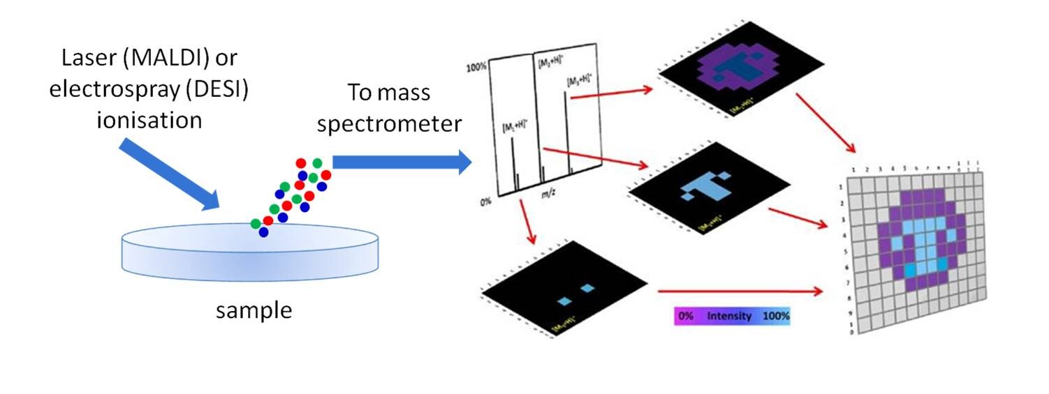
At the American Society for Mass Spectrometry (ASMS) conference in San Antonio (June 6-10), Waters’ scientists and our collaborators demonstrated the utility of MS imaging across a range of applications such as proteomics, metabolomics, cell and tissue biology, research pathology, and histology, and showcased the latest technical developments in these exciting technologies.
Here are our featured posters:
- DESI Imaging at Varying Acquisition Rates with Real Time Imaging Display for Optimized Tissue Imaging
- Automating High Throughput 3D Imaging Using DESI Mass Spectrometry
- Investigation into the Use of Tissue Washing Procedures and the Subsequent Outcomes for DESI-MS Imaging Analyses
To learn more about the power of MS imaging and ion mobility, check out the latest video series by Jose Castro-Perez, Ph.D., director of Health Sciences Marketing for Waters, where he presents a thorough overview of this rapidly growing technique and how it is being used in the mass spectrometry, biomedical, and pathology scientific communities.
Related posts

How Improved Selectivity in Immunosuppressant Analysis Can Help Organ Transplant Patients
The Waters MassTrak Immunosuppressant Calibrator, Control, and Internal Standard Sets, soon available, are developed to help laboratories utilize LC-MS/MS with confidence in their assay results.

The Future Generation of LC-MS/MS IVD Solutions
Our MassTrak IVD solutions provide customers with a range of instrumentation, reagent kits and informatics that can be configured to support laboratory workflows ensuring regulatory compliance.

The Waters Difference: Eliminate Doubt with Chemistry Technical Services
The Waters Chemistry Technical Service Group is made up of world-class chromatography experts with an ability to uncover ways that labs can be made more robust and reliable.
Popular Topics
ACQUITY QDa (16) bioanalysis (11) biologics (14) biopharma (26) biopharmaceutical (36) biosimilars (11) biotherapeutics (16) case study (16) chromatography (14) data integrity (21) food analysis (12) HPLC (15) LC-MS (21) liquid chromatography (LC) (19) mass detection (15) mass spectrometry (MS) (54) method development (13) STEM (12)