A Virus Gives Up One of Its Long-Held Secrets to Researchers at the National University of Singapore

By breaking the Dengue virus into fragment peptides and then digitally reassembling it, scientists identify its vulnerable hot spots
We had a cold, wet spring in the northeastern United States this year. With that rain came standing water, and the standing water was a breeding ground for this summer’s bumper crop of mosquitoes.
For us New Englanders, mosquitoes are mostly a nuisance; for residents of other parts of the world, however, mosquitoes bring disease and death.
One mosquito-borne disease responsible for thousands of deaths, and tens of millions of infections each year, is Dengue fever. One of a number of infectious diseases carried by mosquitoes, it’s a leading cause of illness and death in Puerto Rico, Latin America, Southeast Asia, and the Pacific Islands, according to the United States Centers for Disease Control (CDC). When caught early in patients, prompt treatment can substantially lower the risk of medical complications and death.
One solution for controlling Dengue fever is to eradicate all mosquitoes. Given there are 3,500 species of mosquito it’s an idea that may not work. One Singaporean scientist has another, more practical, idea and his team’s research is opening up new paths of discovery.
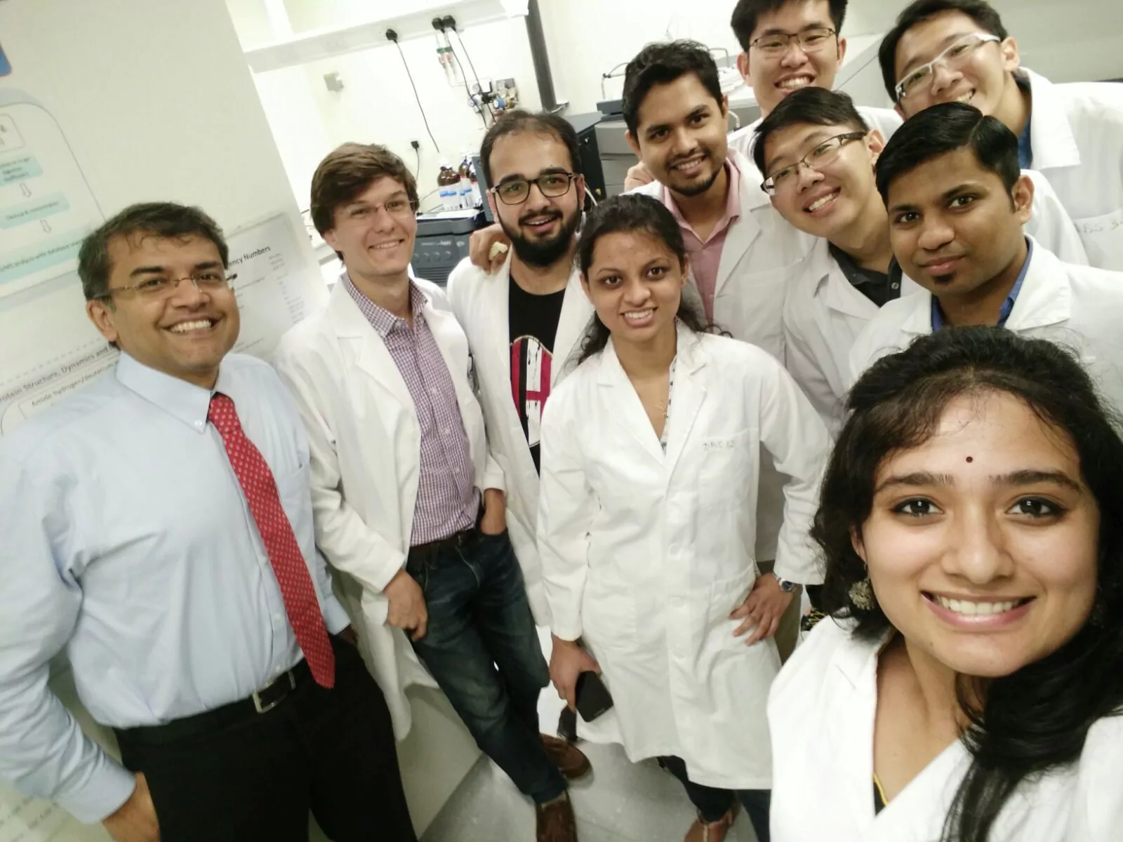
Dr. Ganesh Anand is a structural biologist and an associate professor in the Department of Biological Sciences at the National University of Singapore – a country well-acquainted with Dengue fever. So when Dr. Anand’s research team published papers in Nature Communications and Structure they stirred excitement among infectious disease researchers by revealing something new about viruses that hadn’t been understood before.

Dengue fever: An endemic disease of the tropics yields a secret
“Dengue fever is prevalent here in Singapore. I’ve never contracted it but I know students who have. In India, where I grew up, and in the rest of the tropics, it’s a huge problem. If you recover from the initial infection, it usually doesn’t cause any lasting problems. What you have to watch out for are the second infections, which can be life-threatening,” Dr. Anand says.

Several strains, or serotypes, of the Dengue virus exist in nature but they tend to share one thing in common. “If you do a Google search for the virus, the images of the viral particles look remarkably similar,” he says. “They all look like very rigid structures, almost like very compact, soccer ball-like arrangements.”
One question that has always intrigued Dr. Anand is how does a seemingly rigid particle like a virus that has no sensory organs like eyes and ears, realize when it has entered a human host and know it’s time to enter its infectious phase?
To his surprise, and fascination, the answer to that question is: temperature.
When an infected mosquito bites its victim, the mosquito transmits the Dengue virus in its salivary glands into the bloodstream of its host and that starts an incubation period of several days during which the virus begins replicating.
“The temperature of a cold-blooded mosquito is 28 degrees Centigrade or lower. At that temperature, we now know that the virus assumes a very compact, uniformly rigid shape approximately 500 angstroms in size. But once the virus enters its human host, it reaches 37 degrees Centigrade and takes on a larger, rougher, non-uniform shape exposing all these gaps on its surface,” says Dr. Anand. “To our amazement, they are not rigid rocks after all, but very dynamic entities. For us, that was a big ‘aha’ moment.”
That swelling or expansion of the virus capsid is irregular and the gaps, or vulnerabilities, that appear on the three-dimensional surface aren’t that easy to spot, even for the most powerful microscopes. That’s where ion mobility hydrogen-deuterium exchange mass spectrometry comes in.
‘Visualizing’ a virus particle’s surface and hot spots with mass spectrometry
No stranger to mass spectrometry, Dr. Anand has pioneered one technique that has proven valuable for characterizing not only how molecules are put together, but how they are shaped.
“We had done a lot of previous work on harmless bacteriophages using ion mobility hydrogen-deuterium exchange mass spectrometry (HDX-MS). We knew how valuable it is for studying bacteriophage particle dynamics. So before we decided to use it on the Dengue virus, we knew what to expect and what challenges we were likely to encounter,” he said.
Dr. Anand explains how HDX works: “We take the viral particle and blast it into small pieces with pepsin, and then track how much deuterium is exchanged for hydrogen by the fragment peptides. Each of these peptides represent reporters, which tell you where the expansion is occurring the most and where the action is. With the data processing software we can reassemble the virus particle and accurately pinpoint where on the surface most hydrogen-deuterium exchange occurs. These sites represent possible vulnerabilities or targets. We call them HDX hot spots,” he said.
“Once we have that information, we can overlay it onto the structure of the virus and pinpoint the hotter regions, or those with a higher increase in the deuterium exchange. So the peptides are really great reporters for studying whole viral particle dynamics and doing any kind of perturbation analysis. We are now seeing another dimension to how a virus breathes in solution.”

With a challenging sample, mass spectrometry sensitivity makes a difference
As Dr. Anand explains, viruses are tricky to work with. “One major constraint in working with viruses is that they tend to resist being super concentrated and become aggregated.
“Mass spectral sensitivity is a prerequisite for our studies and the Waters SYNAPT G2-Si mass spectrometer proved fantastic and critical for us to be able to visualize viruses by HDX because of its exquisite sensitivity – we never really had to get to very high concentrations of viral particles in solution. For this study, we used dengue virus particles at a concentration equivalent to 0.25 mg/mL of the envelope E-protein, 180 copies of which form the envelope of each virus particle,” he said.
“If it weren’t for the SYNAPT G2-Si, we couldn’t have done what we did, so I just want to emphasize how critical it is to have such a high-sensitivity instrument to visualize viruses by HDX.” – Dr. Ganesh Anand
What’s next for Dr. Anand’s unlocked Dengue virus?
“So, now we can begin to mine some of these hot spots for designer therapeutics or antibodies. We have recently published the epitope and paratope of a neutralizing antibody that finds the envelope protein of Dengue virus. The powerful aspect of this story is that we have captured how the antibody engages the entire viral particle,” he said.
“What is fascinating is that the antibody appears to be flexible enough to engage with the dengue viral particle and its dynamic movements as the virus changes in response to temperature. Further, the heavy chain alone of the antibody was sufficient for engaging and neutralizing the dengue virus. This presents a lot of exciting opportunities for engineering newer antibodies. It has all kinds of antiviral therapeutic implications.”
Dr. Anand’s team’s papers in Nature Communications – and now Structure – have stirred a lot of scientific discussion and interest. “We’ve received a lot of positive feedback from all over the world. Wherever I’ve given talks, the virologists and immunologists in the audience are quite amazed and excited – I mean, no one associates mass spectrometry with this level of visualization. They’re extremely aware of the implications for targeting viruses of all kinds,” he said.
“So, for us the challenge is to demonstrate that, yes, we can mine these hot spots, and possibly target them with neutralizing antibodies or other therapeutics. That’s the challenge that keeps me up at night.”
Popular Topics
ACQUITY QDa (16) bioanalysis (11) biologics (14) biopharma (26) biopharmaceutical (36) biosimilars (11) biotherapeutics (16) case study (16) chromatography (14) data integrity (21) food analysis (12) HPLC (15) LC-MS (21) liquid chromatography (LC) (19) mass detection (15) mass spectrometry (MS) (54) method development (13) STEM (12)


